


The genus
Specimens of
For identification, each specimen was examined for morphological characteristics such as colony, system, stigmata, stomach, gut, gonad and coloration under stereomicroscope (SMZ 745T; Nikon, Tokyo, Japan). The color of each part was recorded in accordance to a color chart (Pantone color formula guide 747XR). Images of the collected living
The systematic scheme of ascidians was adopted from Kott (1992), Monniot (2001), and Sanamyan (2015). The specimens were deposited in Natural History Museum, Ewha Womans University, Seoul, Korea (EWNHMAS 13-17).
>
Material examined. Korea: Jeju-do: 4 colonies, Munseom, 7 Nov 2000, 15-25 m deep by SCUBA diving (EWNHMAS 13); 1 colony, Beomseom, 6 Jun 2001, 25 m deep by SCUBA diving (EWNHMAS 14); 3 colonies, Munseom, 27 Nov 2011, Song JI, Whang SJ, 20-25 m deep by SCUBA diving (EWNHMAS 15); 2 colonies, Munseom, 28 Nov 2011, Song JI, Whang SJ, 20 m deep by SCUBA diving (EWNHMAS 16); 1 colony, Beomseom, 28 Oct 2014, Whang SJ, Lee WK, 13-14 m deep by SCUBA diving (EWNHMAS 17).
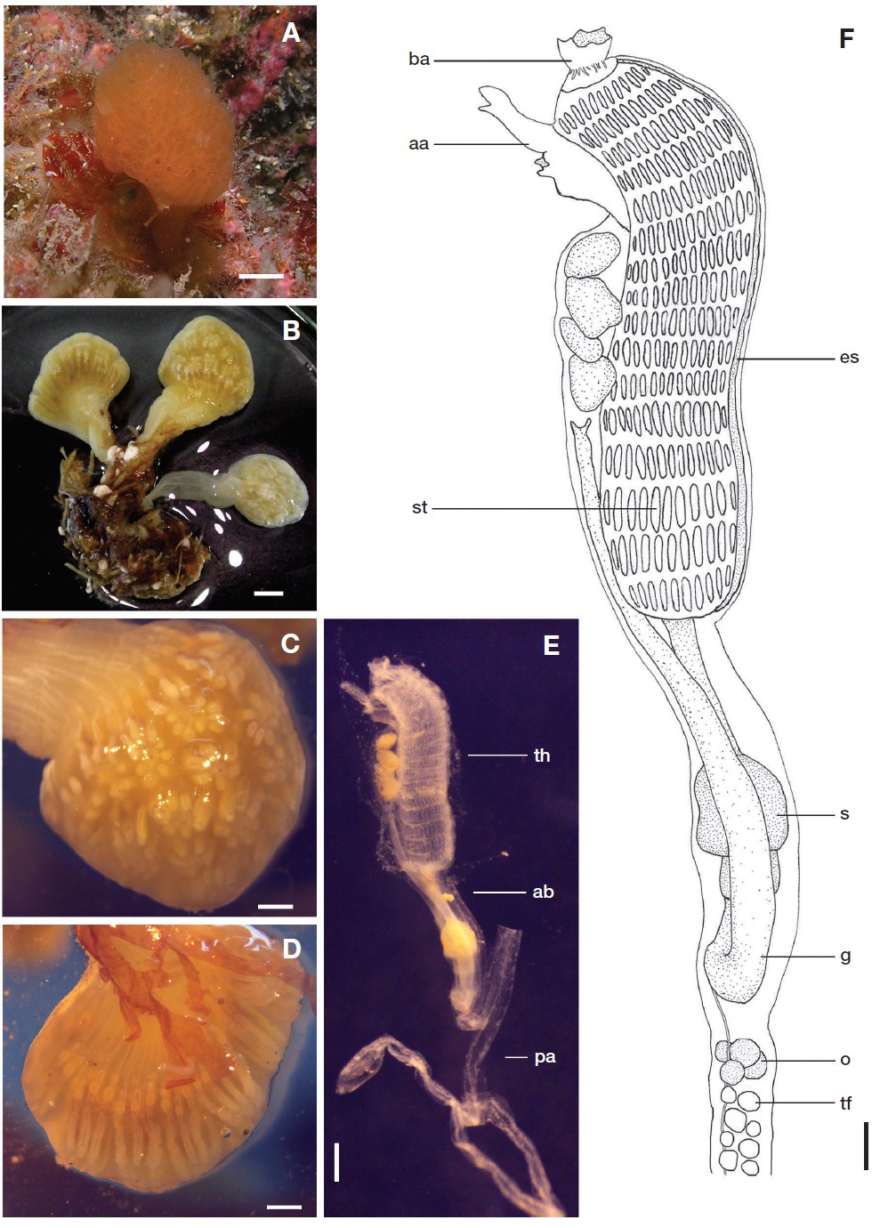
Description. Colonies attached to substrate, consist of several club-shaped cormidia united one another at basal end. Each of cormidia comprises oval head (9.40-22.00 mm in diameter, 6.36-20.00 mm in length) and elongate stalk (up to 20 mm). Surface of colony smooth, sometimes bryozoa and hydra lived on stalk. Test soft, gelatinous, semi-transparent, light orange (Pantone 1375 C) in living and opaque milky white color in preservative (Fig. 1A, B).
Five to eleven zooids form a system encircling a small common cloacal aperture. Eight to thirty systems in oval head of each cormidium (Fig. 1C). Many zooids arranged parallel to axis of cormidium (Fig. 1D).
Zooids deep orange color (Pantone 1585 C) in living (Fig. 1A). Zooids slender and 9.44-16.40 mm in total length in preserved materials. Thorax (2.93-4.12 mm) longer than abdomen (1.63-2.67 mm). Post-abdomen 4.02-10.43 mm in length. Zooids lack constriction between abdomen and post-abdomen. Thin vascular canal is issued from posterior end of post-abdomen and reach deep into stalk. About 10 thin thoracic longitudinal muscles on each side of thorax. Branchial aperture has six lobes and atrial aperture has a slender languet. Branchial tentacles 16-30. Branchial sac has 14-16 stigmata rows of 10-17 stigmata (or rarely 20 stigmata) per row. Dorsal languet fine pointed tongue shape. Stomach yellow, elliptical, have smooth surface without longitudinally folded wall and situated nearly in middle of abdomen. Mid-intestine distinct and has no coecum. Anus opens at level as high as posterior fifth stigmata row and bilobed (Fig. 1E, F).
Several ovaries situated just behind intestinal loop and small testicular follicles arranged densely in post-abdomen of Munseom’s sample collected on 7 Nov 2000.
Distribution. Pacific Ocean: Korea (Beomseom and Munseom, Jeju-do), Japan (Pacific coast of Honsyu; Shiranezaki, Isl. Dogo; Sagami Bay; off Wakayama Pref).
Remarks. This is the first record of
The present specimens resemble Tokioka’s (1954) redescription of
Korean name:1*곤봉만두멍게 (신칭)