검색 전체 메뉴
PDF
맨 위로
OA 학술지
Structural and thermal characteristics of photocrosslinked silk fibroin - PEG hydrogel
  • 비영리 CC BY-NC
  • 비영리 CC BY-NC
ABSTRACT
Structural and thermal characteristics of photocrosslinked silk fibroin - PEG hydrogel
KEYWORD
silk fibroin , poly(ethylene glycol) , hydrogel , sonication , photopolymerization
  • Introduction

    Hydrogels are crosslinked hydrophilic matrices suitable for a variety of biomedical applications, including tissue engineering matrix, contact lenses, drug delivery carriers and cell therapy (Kweon et al., 2014; Lin and Anseth, 2009; Kweon et al., 2001; Hanawa et al., 1995). A variety of natural and synthetic polymers have been explored as the base materials for hydrogel crosslinking. Silk fibroin (SF) produced by Bombyx mori silkworm can be formulated into physical hydrogels or be modified to display reactive motifs (e.g., methacrylate) for chemical crosslinking (Yecel et al., 2014; Kim et al., 2014; Kasoju and Bora, 2012; Kweon et al., 2001; Kweon et al., 2000). SF can also form β-sheets structure, which can self-assemble into higher order structures with enhanced stability. On the other hand, hydrogels prepared from derivatives of poly(ethylene glycol) (PEG) chemically crosslinked with limited degradability (Lin, 2015; Lin and Anseth, 2009; Lutolf and Hubbell, 2005).

    Among the various physical and chemical methods for hydrogel crosslinking, we are particularly interested in visible light induced gelation (Hao and Lin, 2014; Hao et al., 2014; Kweon et al., 2001). Especially, thiol-acrylate photopolymerization not only permits rapid and efficient crosslinking, but also yields hydrolytically degradable hydrogels without the need to synthesize degradable macromers (Hao and Lin, 2014; Hao et al., 2014).

    Gelation of SF depends on a number of parameters including concentration, temperature, mechanical strength, chemical treatment, ultrasound sonication, and composition (Kim et al., 2015; Samal et al., 2013; Wang et al., 2008; Kweon et al., 2000; Kang et al., 2000; Ayub et al., 1994). Among them, sonication induced rapid gelation by reduction of molecular weight and β-sheet conformation (Samal et al., 2013).

    The authors reported the modulating properties of SF-PEG hydrogels were reported (Bragg et al., 2016). In this study, we examined the structural characteristics of SF-PEG hydrogel. Aqueous SF was mixed with PEG macromer solution containing PEG-diacrylate (PEGDA), dithiothreitol (DTT), and N-vinylpyrrolidone (NVP) to form hydrogel. Visible light mediated thiol-acrylate photopolymerization was performed to make SF hydrogel with chemically crosslinked and hydrolytically labile thiol-acrylate. We also examined the effect of sonication on the SF solution on the SF-PEG hydrogel.

    Materials and methods

      >  Materials

    SF was purified from Bombyx mori silkworm as described previously (Kim et al., 2014; Kweon et al., 2008). PEGDA (3.4kDa) was synthesized following an established protocol (Hao and Lin, 2014). Eosin-Y disodium salt was purchased from MP Biomedical and used without purification. All other chemicals and reagents were obtained from Thermo Fisher Scientific unless noted otherwise.

      >  Preparation of SF aqueous solution

    SF was dissolved in ethanolic calcium chloride solution (CaCl2 : H2O : ethanol = 1 : 8 : 2 in molar ratio). The solution was heated to 90℃ for 4 h, cool down to room temperature, and subsequently dialyzed against ddH2O using dialysis membrane with MWCO 6-8 kDa (Fisher) for 2 d to remove the salts. After dialysis, the SF solution contained in the dialysis membrane was placed in a bath of dry PEG (10kDa) to concentrate the SF solution. The final concentration (wt./vol.%) of SF aqueous solution was determined from lyophilization of a small portion of the dialyzed SF solution (5 – 6 wt%). Sonication of SF solution was achieved using a Branson 450 Sonifier with a converter, an externally threaded disruptor horn, and 1/8” diameter-tapered microtip. To avoid forming, SF solution was sonicated for 1 s at 10% amplitude and then sonicated at 20% amplitude for 20 s (Wang et al., 2008). The silk was used for hydrogel formation within 30 min after sonication.

      >  Hydrogel formation initiated by visible light

    Hydrolytically degradable PEG hydrogels were formed by visible light initiated thiol-acrylate photopolymerization using PEGDA and dithiol liker. A typical pre-polymer solution was prepared in pH 7.4 PBS and contained macromer PEGDA (10 wt%), photosensitizer eosin-Y (0.1 mM), bi-functional co-initiator DTT (7.5 mM), and co-monomer NVP (0.1 vol%). macromers (Hao and Lin, 2014; Hao et al., 2014). All concentrations indicated were final concentrations in the pre-polymer solutions. Aliquots of the pre-polymer solution were subjected to halogen cold light (400-700 nm, AmScope, Inc.) exposure for 5 min (10mW/cm2 at 550nm or 70k Lux) between glass slides with spceres. In some experiments, soluble SF was added at different weight contents.

      >  Instrumental analysis

    Surface morphology of SF hydrogel was observed using scanning electron microscope (S-3500N, Hitachi, Japan). The hydrogels were coated with platinum and before observation. To investigate the molecular structure of the hydrogels, X-ray diffraction (XRD) was measured on X’Pert-Pro (PANalytical, Netherlands) using Cu-Kα radiation. The X-ray source was operated at 40 kV and 40 mA. Differential scanning calorimetry was analyzed using Differential Scanning Calorimeter (Mettler Toledo, Australia) at a scanning rate of 10℃/min over 50-350℃. Themrogravimetric analysis was done using Mettler Toledo ThermoGravimetric Analyzer (DSC 823e, Australia) from 35℃ to 800℃ at the heating rate 10℃/min with a nitrogen flow rate of 50 mL/min.

    Results and discussion

    In this study, SF-PEG hydrogel crosslinked from PEGDA, co-monomer NVP, di-thiol containing linker DTT, and photosensitizer eosion-Y were evaluated by instrumental analysis on morphological, structural, and thermal properties.

      >  Morphology of SF-PEG hydrogel

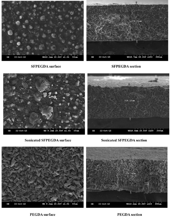
    To observe the surface and fractured section of SF hydrogel, SEM is a powerful method. As Fig. 1, Surface of SFPEGDA showed dot and uneven surface morphology with network cross-section structure. Sonicated SFPEGDA shows some big and small islets with network cross section. However, PEGDA showed that relatively even surface morphology with network structure. This results means that SF and PEGDA were phase-separated and results in that SF was distributed in the PEG matrix.

      >  Crystalline structure

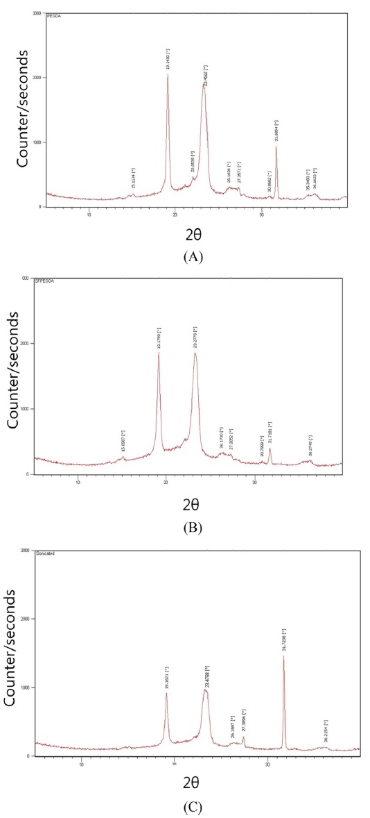
    XRD curves of SF-PEG hydrogels are shown in Fig. 2 SF showed a typical amorphous diffraction pattern (data not shown), whereas PEGDA hydrogel showed sharp diffraction peak at 2θ=19.1, 23.5, and 31.7° (Kweon et al., 2001; Priola et al., 1993). The diffraction pattern of SFPEG (Fig. 2(B)) is consistent with that of the PEGDA itself, regardless of SF entrapment. However, by sonication, the relative diffraction intensities at 2θ= 31.7° and 27.4° were increased.

    SF was entrapped in crosslinked PEG networks. XRD curves show that entrapped SF does not affect the crystal structure of PEG. Kweon et al. (2001) reported that the diffraction pattern of PEGDA did not changed by semi-interpenetrating polymer networks composed of SF and PEG. Liang et al. (1995) also reported that the crystalline structure of PEG did not changed by blending with cellulose. However, some diffraction peaks were changed by sonication; it means that the crystalline structure was affected by molecular agitation induced by sonication.

      >  Thermogravimetric analysis

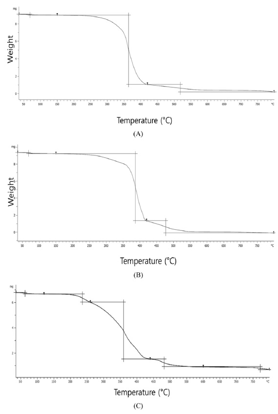
    Fig. 3 showed the thermogravimetric curves of PEGDA, SFPEGDA and sonicated SFPEGDA. PEGDA showed slightly two step mass loss around 230-350℃ and 350-390℃. SFPEGDA showed two step weight losses around 220-360℃ and 360℃ and 390℃. On the other hand, sonicated one showed more complex degradation behavior from 180-420℃

    Decomposition of pure PEG were occurred in one step around 270-370℃ (Massoumi et al., 2015). The PEGDA with acrylate group decomposed around 230-350℃ and followed around 350- 390 ℃ with the main chain scission of PEG. On the other hand, SFPEGDA showed more clear two step degradation behaviors with amino acid composition containing hydrophilic side group and main chain scission. As shown in Fig. 1, sonication induced some aggregation of molecules, therefore sonicated one showed more complex degradation pattern

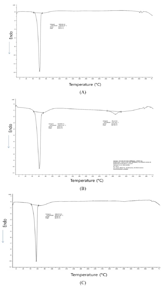
    Fig. 4 showed the DSC thermograms of PEGDA, SFPEGDA, and sonicated SFPEGDA. PEGDA shows a sharp endothermic peak at 59.2℃ with ΔHm 128.6 J/g and SFPEGDA shows two endothermic peaks at 60.2℃ with 108.2 J/g and at 287℃ with 9.5 J/g. Sonicated SF hydrogel (Fig. 4(c)) also shows two endothermic peaks at 55.8℃ with 99.6 J/g and weak peaks around 290℃.

    The melting temperature of PEG crystalline observed at 50- 60℃ (Icart et al., 2016; Kweon et al., 2001). So, the sharp endothermic peak around 60℃ in Fig. 4, Tm of PEG crystalline region, was slightly shifted to higher temperature by entrapment with SF. The degree of crystalline is related to the melting enthalpy ΔHm. The melting enthalpy of SFPEGDA 108.2 J/ g was lower than that of PEGDA, means that the degree of crystalline was decreased with a decrease of melting enthalpy. By sonication, the degree of crystalline region of PEG was significantly decreased. On the other hand, the endothermic peak around 287℃ is considered as the degradation of SF itself. Many researchers reported that SF degraded around 270-380℃ with structural characteristics (Kweon et al., 2001; ). These results are corresponding with the TGA results (Fig. 3).

참고문헌
  • 1. Ayub ZH, Arai M, Hirabayashi K (1994) Quantitative structural analysis and physical properties of silk fibroin hydrogels. [Polym] Vol.35 P.2197-2200 google cross ref
  • 2. Bragg JC, Kweon HY, Jo YY, Lee KG, Lin CC (2016) Modulating properties of chemically crosslinked PEG hydrogels via physical entrapment of silk fibroin. [J Appl Polym Sci] Vol.132 P.43705 google
  • 3. Hanawa T, Watanabe A, Tsuchiya T, Ikoma R, Hidaka M, Sugihara M (1995) New oral dosage form from elderly patients: preparation and characterization of silk fibroin gel. [Chem Pharm Bull] Vol.43 P.284-288 google cross ref
  • 4. Hao Y, Lin CC (2014) Degradable thiol-acrylate hyrogels as tunable matrics for three-dimensional hepatic culture. [J Biomed Mater Res A] Vol.102 P.3813-3827 google cross ref
  • 5. Hao Y, Shih H, Munoz Z, Kemp A, Lin CC (2014) Visible light cured thiol-vinyl hydrogels with tunable degradation for 3D cell culture. [Acta Biomater] Vol.10 P.104-114 google cross ref
  • 6. Icart LP, dos Santos ERF, Pereira ED, Ferreira SR, Saez V, Ramon JA, Nele M, Pinto JCS, Toledo RD, Silva DZ, Souza FG (2016) PLA-b-PEG/magnetite hyperthermic agent prepared by Ugi four component condensation. [eXPRESS Polym Let] Vol.10 P.188-203 google cross ref
  • 7. Kang GD, Nahm JH, Park JS, Moon JY, Cho CS, Yeo JH (2000) Effects of poloxamer on the gelation of silk fibroin. [Macromol Rapid Commun] Vol.21 P.788-791 google
  • 8. Kasoju N, Bora U (2012) Silk fibroin in tissue engineering. [Adv Healthc Mater] Vol.1 P.393-412 google cross ref
  • 9. Kim SK, Jo YY, Lee KG, Lee HS, Yeo JH, Kweon HY (2014) Preparation and characterization of silk beads for protein delivery system. [Int J Indust Entomol] Vol.28 P.66-70 google cross ref
  • 10. Kim SK, Jo YY, Lee KG, Kim KY, Kim HB, Kweon HY (2015) Effect of ethanol concentration on the infrared spectroscopic characterization of silk beads. [J Seric Entomol Sci] Vol.53 P.118-123 google
  • 11. Kweon HY, Jo YY, Lee KG, Kim HB, Yeo J (2014) Silk polymer for medical applications. [J Seric Entomol Sci] Vol.52 P.89-95 google
  • 12. Kweon HY, Yeo JH, Lee KG, Lee HC, Na HS, Won YH, Cho CS (2008) Semi-interpenetrating polymer networks composed of silk fibroin and poly(ethylene glycol) for wound dressing. [Biomed Mater] Vol.3 P.034115 google cross ref
  • 13. Kweon HY, Park SH, Yeo JH, Lee YW, Cho CS (2001) Preparation of Semi-interpenetrating polymer networks composed of silk fibroin and poly(ethylene glycol) macromer. [J Appl Polym Sci] Vol.80 P.1848-1853 google cross ref
  • 14. Kweon HY, Yeo JH, Lee KG, Lee YW, Park YH, Nahm JH, Cho CS (2000) Effects of poloxamer on the gelation of silk sericin. [Macrom Rapid Commun] Vol.21 P.1302-1305 google
  • 15. Liang XH, Guo YQ, Gu LZ, Ding EY (1995) Crystalline-amorphous phase-transition of a poly(ethylene glycol) cellulose blend. [Macromolecules] Vol.28 P.6551-6555 google cross ref
  • 16. Lin CC, Anseth KS (2009) PEG hydrogels for the controlled release of biomolecules in regenerative medicine. [Pharm Res] Vol.26 P.631-643 google cross ref
  • 17. Lin CC (2015) Recent advances in crosslinking chemistry of biomimetic poly9ethyelene glycol) hydrogels. [RSC Adv] Vol.5 P.39844-39853 google cross ref
  • 18. Lutolf MP, J. A. Hubbell JA (2005) Synthetic biomaterials as instructive extracellular microenviroments for morphogenesis in tissue engineering. [Nat Biotechnol] Vol.23 P.47-55 google cross ref
  • 19. Massoumi B, Ramezani m, Jaymand m, Ahmadinejad M (2015) Multi-walled carbon nanotubes-g-poly(ethylene glycol)-b-poly(e-caprolactone): synthesis, characterization, and properties. [J Polym Res] Vol.22 P.214 google cross ref
  • 20. Priola A, Gozzelino G, Ferrero F, Malucelli G (1993) Properties of polymeric films obtained from UV cured poly(ethylene glycol) diacrylates. [Polymer] Vol.34 P.3653-3657 google cross ref
  • 21. Samal SK, Kaplan DL, Chiellini E (2013) Ultrasound sonication effects on silk fibroin protein. [Macromol Mater Eng] Vol.298 P.1201-1208 google cross ref
  • 22. Yucel T, Lovett ML, Kaplan DL (2014) Silk-based biomaterials for sustained drug delivery. [J Control Release] Vol.190 P.381-97 google cross ref
  • 23. Wang X, Kluge JA, Leisk GG, Kaplan DL (2008) Sonication-induced gelation of silk fibroin for cell encapsulation. [Biomaterials] Vol.29 P.1054-64 google cross ref
OAK XML 통계
이미지 / 테이블
  • [ Fig. 1. ]  SEM photographs of SF-PEG hydrogels.
    SEM photographs of SF-PEG hydrogels.
  • [ Fig. 2. ]  X-ray diffraction patterns of PEGDA(A), SFPEGDA(B), and sonicated SFPEGDA(C).
    X-ray diffraction patterns of PEGDA(A), SFPEGDA(B), and sonicated SFPEGDA(C).
  • [ Fig. 3. ]  Thermogravimetric curves of PEGDA(A), SFPEGDA(B), and sonicated SFPEGDA(C).
    Thermogravimetric curves of PEGDA(A), SFPEGDA(B), and sonicated SFPEGDA(C).
  • [ Fig. 4. ]  Differential calorimetric curves of PEGDA(A) and SFPEGDA(B) and sonicated SFPEGDA(C).
    Differential calorimetric curves of PEGDA(A) and SFPEGDA(B) and sonicated SFPEGDA(C).
(우)06579 서울시 서초구 반포대로 201(반포동)
Tel. 02-537-6389 | Fax. 02-590-0571 | 문의 : oak2014@korea.kr
Copyright(c) National Library of Korea. All rights reserved.